Concept
Bronchopneumonia is very common among young animals. The disease has a second name - catarrhal pneumonia. A large number of calves that become ill with bronchopneumonia die. Accordingly, serious financial damage is caused to the economy.
The disease affects the respiratory system of the calf's body. In particular, the alveoli and bronchi are subject to inflammation. Sometimes bronchopneumonia affects other organs. If timely treatment measures are not taken, intoxication of the entire body, which has not yet become stronger, occurs.
Bronchopneumonia can be cured. But after suffering from an illness, consequences occur:
- Calves do not gain weight well.
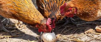
- The disease affects the breeding qualities of bulls.
- Reproductive qualities deteriorate.
The disease is more likely to be a cold than an infectious disease. She's not contagious. It can occur from a severe cold or hypothermia.
Bronchopneumonia does not develop in any specific region or area. It is widespread everywhere. In terms of the number of registered cases, it ranks second after gastrointestinal disorders.
Calves between 30 and 45 days of age are most susceptible to bronchopneumonia. The percentage of cases among young animals is approximately 30% of the entire population.
Causes
The etiology of the disease is quite diverse. Let's consider the main reasons for the development of bronchopneumonia in young animals:
- Crowded housing of animals.
- Contaminated air.
- Poor quality of nutrition (lack of vitamins and amino acids in the diet).
- Unsuitable living conditions for animals (unheated room, dampness, lack of bedding).
- Stressful situations.
- Hypothermia.
- Congenital pathologies - short trachea, narrow lumens of the bronchi, saturation of the mucous membrane of the respiratory tract with capillaries, etc.
Poor food quality
Under the influence of unfavorable factors, immunity decreases and pathogenic bacteria - streptococci, staphylococci, E. coli, Pseudomonas aeruginosa, pneumococci, fungi, etc. - begin to rapidly multiply in the bronchi of the calf. This leads to general intoxication of the body and destructive changes in the organs of the respiratory system.
Disease development process
The disease develops over a long period of time. Initially, bronchopneumonia affects the bronchi and spreads along the bronchial tree. Treatment of the developed chronic form of the disease causes certain difficulties, because a serious inflammatory process occurs, accompanied by high fever and swelling of the mucous membranes in the nasal passages.
After a few days, signs of inflammation in the lungs and wheezing appear.
Depending on the severity and severity of the disease, symptoms may increase or decrease. The acute form is usually accompanied by swelling of the lungs and pallor of the mucous membranes.
Bronchopneumonia is always accompanied by a cough, since the tissues become denser at the sites of inflammation in the lungs. Coughing is a natural reaction of the body.
Why does a calf or cow cough: what to do and how to treat it
Young animals are more susceptible to respiratory diseases. One of the manifestations of these ailments is cough. If a cow is coughing, the farmer needs to find the cause of the disease as quickly as possible in order to begin treatment on time and avoid complications.
Causes of cough
Cough in calves can occur for various reasons. For example, if young animals are kept in an unheated room on a cement floor without bedding. Hypothermia is a factor that contributes to the development of the inflammatory process in the bronchi and lungs.
The humidity in the room is also important. Dust, the presence of harmful vapors in the air - ammonia or hydrogen sulfide, can cause coughing in young animals
A stressful situation can also provoke a decrease in immunity and inflammation in the respiratory tract:
- Transportation of animals.
- Weaning from mother.
- Castration.
Infectious and parasitic diseases pose the greatest danger. They always occur against the background of decreased immune defense. These include:
- Tuberculosis.
- Inflammation of the lungs - pneumonia, as well as bronchopneumonia.
- Dictyocaulosis.
These ailments are especially susceptible to young individuals, whose immunity is still weak enough to resist viruses and bacterial infections. Animals that are rarely outdoors and whose diet is poor are much more likely to get sick.
Calves become infected with infectious diseases during grazing, through contact with sick individuals, through water, soil and food, as well as from the mother through milk. Let's look at how these ailments manifest themselves.
Infection from mother through milk
Symptoms
If a calf has become infected with an infection, one of the manifestations of which is coughing, then this can be determined by the following symptoms:
- The animal is weakened and depressed.
- The cough is dry, frequent or wet.
- The temperature rises to 40-41 degrees.
- Mucus is discharged from the nose.
- In some diseases, pallor of the mucous membranes is noted.
- Tuberculosis and bronchopneumonia are often accompanied by diarrhea.
- No appetite.
- Shortness of breath appears.
- Wheezing can be heard.
- With tuberculosis, the lymph nodes located under the jaw and on the neck usually become enlarged.
Having noticed at least a few symptoms in a calf, in addition to coughing, the farmer needs to immediately call a veterinary service worker. Treatment cannot be started without establishing an accurate diagnosis.
Attention! If a calf is coughing, it should be separated from the herd to avoid spreading infection.
Diagnostics
When examining a calf, the veterinarian takes into account the body temperature of the sick cow, examines the mucous membranes, and listens to the lungs and heart.
Although these data help to navigate and guess what the animal is sick with, the results of laboratory tests are still important. Many diseases at the initial stage of development are similar; to avoid errors in diagnosis, it is recommended to take blood samples, mucus samples, and also take an x-ray of the lungs
Many diseases at the initial stage of development are similar; to avoid errors in diagnosis, it is recommended to take blood samples, mucus samples, and also take an x-ray of the lungs.
The image shows changes in the form of darkening, having either clear contours or blurred ones, multiple or single foci of inflammation are differentiated
The veterinarian pays attention to how the border of the heart is visible. This data helps to exclude certain diseases or find out in what form they occur
In some cases, a lung biopsy is used to make a diagnosis.
Based on laboratory data, clinical manifestations and images, the veterinarian excludes diseases that are initially included in the list of suspected ones:
- Chlamydia.
- Pleuropneumonia.
- Viral diarrhea.
- Bronchitis and others.
Next, based on the diagnosis, appropriate therapy is prescribed.
Treatment
The treatment regimen depends on the cause of the cough. A sick calf must be isolated from other animals. He needs to be provided with good living conditions - a warm room, soft bedding, peace, good nutrition and plenty of drink. Let's look at how cough in calves and adult cows is treated for tuberculosis, pneumonia and dictyocaulosis.
Reasons for the development of bronchopneumonia
Usually, the factors causing the appearance of an inflammatory process in the respiratory system are exclusively external reasons, which may deteriorate the immunity of young animals, as well as microbes and bacteria.
The onset of the disease is promoted by:
- High level of air humidity in the area where calves are kept.
- Drafts and cold.
- Heat and dry indoor air.
- Dirty, dusty air.
- Unbalanced feeding.
- Lack of vitamin A and C.
- Violation of colostrum feeding.
- Stressful situations.
- Incorrectly selected pair during crossing.
- Problems with the digestive system.
- Untreated colds.
Preventive actions
Cough is only a symptom of pathologies of the respiratory system (less often than other departments), therefore preventive measures are similar to measures to prevent bronchitis, pneumonia and other diseases.
First of all, regular vaccination is carried out against the most common infections in the region. For this purpose, laboratory diagnostics are carried out for the presence of the pathogen and a specific vaccine is developed. Polyvalent drugs are used more often against RTIs, coronavirus infections, parainfluenza 3, salmonellosis, and enterococcal infections.
Cows and young animals are provided with optimal living conditions
It is important to prevent high humidity on the farm, equip the premises with ventilation, and regularly clean and disinfect. Aerosols should be used as disinfectants to simultaneously prevent lung infections
The temperature in the barn should not fall below 14 degrees and exceed 25. At the same time, beef breeds are less sensitive to temperature and can be kept outdoors in winter, but they must be provided with good nutrition and shelter from precipitation and wind.
Feeding must take into account physiological norms
It is important to ensure the norm for protein, vitamins A and D, since their deficiency leads to disruption of the epithelial covers of the respiratory tract. Farms should carry out routine immunizations
Imported livestock must be subjected to quarantine measures and diagnosed for infectious and invasive diseases (tuberculosis, dictyocaulosis). The herd should be formed from strong calves, free from hidden and chronic diseases.
Acute
It develops in the body in a little over a week. Requires immediate veterinary examination and collection of tests for the presence of infection and detection of leukocytosis in the blood and protein in the urine.
Symptoms:
- lethargy;
- poor appetite;
- throat breathing;
- lacrimation;
- mucus secreted from the nasal passages;
- the appearance of a cough accompanied by unnatural sharp sounds.
If drug treatment measures are not taken in time, the symptoms of the disease will rapidly begin to intensify. The cough turns from dry to wet and more frequent.
Causes of pneumonia in cattle
It is customary to identify the following reasons why young animals develop pneumonia:
- Unfavorable living conditions. If calves sleep on a cold, damp floor without bedding and are in a room that is rarely ventilated, the risk of pneumonia increases greatly.
- Stress after early weaning. It is not recommended to wean young calves from their mother's milk too early.
- Castration in young bulls.
- Poor quality feed or poor diet. In particular, calves often develop pneumonia during the transition from milk feeding to roughage, since any sudden change in diet weakens the animal's body.
- Illiterate transportation. Animals can catch a cold during transportation in the cold season.
- Lack of movement and oxygen deficiency due to rare walking. Without movement, the animals' muscles atrophy, as a result of which the ventilation of the lungs weakens.
- Viral and bacterial infections.
- Overheating in the sun, which causes disruption of thermoregulation.
- Keeping a large number of animals in cramped conditions. When a large number of cows and calves gather in one room, large amounts of ammonia and hydrogen sulfide quickly accumulate in the air, which does not have the best effect on the health of cattle.
Diagnostics
An accurate diagnosis can be made by conducting a number of laboratory tests:
- X-rays of light.
- Bronchopulmonary test.
- Blood chemistry.
- Analysis of urine.
Any information is important to make a correct diagnosis. Therefore, attention is paid not only to laboratory and clinical studies, but also to the sanitary and hygienic conditions in which the young animals are kept.
The behavior of a sick individual indoors and on a walk is also important.
The research results obtained form the basis for the prescribed treatment.
Treatment costs
Considering the short period of time required to treat a sick individual and the small range of drugs needed for therapy, farms do not incur large financial costs.
A separate topic concerns the weight loss of calves during illness and their slow development in the first period after the illness.
To this will have to be added the costs of repeated calls to the veterinarian and testing.
If young animals die from bronchopneumonia, financial losses will have to be incurred by paying for a pathological examination.
To ensure that the calf breeding business does not suffer losses, you need to take care, first of all, about the health of the young animals. Diseases can destroy numerous herds, resulting in irreparable financial losses.
Such a serious disease as bronchopneumonia is also fatal, but if signs of the disease are detected in a timely manner, it can be quite easily cured.
The main thing is to do everything possible so that the calf recovers from illness as quickly as possible and begins to gain weight. Prevention is the most reliable way to protect against disease.
Changes in the functioning of systems during bronchopneumonia
The mortality rate from bronchopneumonia is very high. This occurs due to late diagnosis or improper treatment. As a result of lung intoxication, malfunctions occur in the functioning of almost all organs and systems.
One of the first to be disrupted is the central nervous system. This provokes:
- decreased protective function of the body;
- blood accumulates in the lungs;
- bronchi and bronchioles swell;
- inflammatory processes appear in the lungs;
- disruption of gas exchange in tissues.
Those areas of the lungs that are already affected by inflammatory processes cease to perform their direct functions, so a greater load is placed on healthy areas. This causes shortness of breath and fever.
When analyzing urine, protein compounds are detected, since bronchopneumonia also affects the kidneys. The operation of the filters in them is disrupted.
The general condition of the gastrointestinal tract and liver plays an important role in the development of the disease. If the liver structure is damaged, then harmful toxins pass through it from the intestines. They are carried throughout the body through the blood and provoke intoxication of internal organs and systems.
What is bronchopneumonia?
This disease is characterized by the development of an inflammatory process in the bronchi and alveoli, accompanied by the accumulation of serous exudate in the bronchial tree. Inflammation quickly spreads, affecting all parts of the respiratory system. The disease always occurs against the background of a general weakening of the immune system, which is why it most often occurs in calves, whose resistance to pathogenic microorganisms is extremely low. The disease is accompanied by general intoxication of the body and can lead to irreversible pathological processes in the organs of the respiratory system, for example, necrosis of lung tissue.
Other causes of cough in calves
Often the cause of cough in calves is the development of completely different pathologies than bronchopneumonia. Therefore, when the first signs of the disease appear, it is worth scheduling an immediate consultation with a veterinarian to clarify and make the correct diagnosis.
Coughing may bother a calf for other reasons:
- entry of a foreign body into the upper respiratory tract;
- pollen ingestion into the respiratory system, which contributes to allergies;
- helminths in the intestines, which cause the accumulation of echinococci in the pulmonary system;
- poisoning with chemical compounds that irritate mucous membranes. Cough in this case is the body’s protective function against an irritant.
Elimination of cough and treatment of the underlying disease
Therapeutic measures differ from the stage of the pathological process. In case of acute and painful cough, its negative impact should be reduced, and in chronic forms it is necessary, on the contrary, to activate resistance and speed up the recovery process.
How to treat the chronic form:
- hemotherapy – intramuscular injections in a dose of up to 0.1 ml/kg. It is recommended to add anti-clotting drugs to the blood, as well as antibiotics. Administration is repeated at daily intervals;
- IR irradiation. The use of infrared light makes it possible to enhance the functioning of internal organs (primarily the heart and lungs), warms the skin, and accelerates metabolic processes. For treatment, cows are irradiated to the chest from a distance of 1-1.5 meters for 15-30 minutes. The procedures are repeated daily until recovery;
- Massaging and rubbing the chest helps increase blood flow. For this purpose, straw strands are used, the procedure is carried out with intense circular movements. To enhance the effect, it is recommended to apply camphor oil to the skin (can be mixed with ichthyol). After the massage, the calf is placed in a warm room and wrapped;
- applications of mud, paraffin, ozokerite, clay. Substances are applied to the skin in a heated state, which leads to a local increase in temperature and local blood flow. The effect intensifies when it dries - the application shrinks, increasing the effusion of blood. The effect is also based on chemical irritation of the skin, some role belongs to the active components of mud or clay.
The action of pathogenetic therapy is aimed at local enhancement of blood circulation. This leads to blood effusion followed by hemolysis, which contributes to an increase in immune status. Cows mobilize the body's protective reserves, this leads to an exacerbation of the old pathogenic process and treatment becomes easier.
At the same time, it is necessary to ensure the separation of exudate from the respiratory tract. The use of steam generators, aerosols and mists shows good results. The mechanism of action is based on the introduction of the drug directly into the affected area. In addition, this technique allows for group treatment activities.
Antibiotics, sulfa drugs and disinfectants are used for spraying. This allows you to destroy the pathogen that has accumulated in the bronchi and upper respiratory tract. To separate the contents, inhalations of turpentine, sodium bicarbonate, pepsin, and vegetable oils (eucalyptus, mint) should be used. In the acute form of the inflammatory process in the respiratory system, coughing causes severe pain. This leads to increased destructive processes and slower recovery. Treatment requires relief of pain; novocaine blockade of the stellate ganglion has a great effect for this purpose:
- cattle are fixed in a lateral position;
- the thoracic limb is retracted as far back as possible;
- find the cranial edge of the first rib;
- the needle is inserted in the upper third of the rib towards the back until it stops at the vertebral body;
- returning the needle a little, inject up to 40 ml of 0.5% novocaine;
- The procedure is carried out on one side only; antibiotics can be added.
It is recommended to provide sick animals with a good diet, regular walks, and keep them in warm, ventilated rooms. In severe cases, compensatory therapy is prescribed. For parasitic diseases, specific agents are used.
Pathological changes
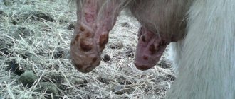
When a calf dies from bronchopneumonia, an autopsy is ordered. During the examination, a number of characteristic changes in the body of a young individual are revealed:
- damage to the bronchopulmonary system;
- compactions in the lungs;
- pneumonia lesions in the upper parts of the lungs, penetrating deep into the organ. The lesions are about 4 centimeters in diameter, red-blue or light gray in color. The surface of the lungs is uneven. When cut, granular formations are observed.
- The bronchi and bronchioles of the animal are filled with dense mucus.
- Lesions of the lymphatic system. In particular, inflammation is observed near the bronchi and lungs.
- Overgrowth of connective tissue.
- Exhaustion of the body.
- Enlarged liver.
- Cardiac muscle of unnatural color.
- Swelling of the upper respiratory tract.
- A bladder full of bile.
Catarrhal bronchopneumonia
Catarrhal bronchopneumonia (Bronchopneumonia catarrhalis) is a lobular inflammation of the bronchi and lungs, accompanied by the formation of catarrhal exudate consisting of epithelial cells, blood plasma, leukocytes and filling the lumen of the bronchi and alveolar cavities.
Catarrhal bronchopneumonia is widespread among animals of all species, causing great economic damage to livestock farming. Most often, young animals suffer from catarrhal bronchopneumonia during weaning, growing and fattening. If veterinary and sanitary rules are violated on large farms, specialized farms and industrial complexes, catarrhal bronchopneumonia can become widespread, affecting up to 30-40% of the total livestock.
Etiology . Catarrhal bronchopneumonia is a polyetiological disease and occurs in animals as a result of the combined effect on the body of unfavorable factors leading to a weakening of resistance. Among the external factors leading to the occurrence of bronchopneumonia, the first place is taken by colds and others associated with irritation of the respiratory tract. These include keeping animals in drafty rooms, high air humidity, hypothermia and overheating, damp floors and walls, lack of bedding material, inhalation of large amounts of dust, ammonia, hydrogen sulfide and other irritants.
In modern industrial complexes and specialized farms for raising heifers and fattening cattle, the main reasons for the occurrence and spread of the disease are various violations committed by farm specialists during the acquisition process, and violations of existing livestock standards: hypothermia of calves during transportation from the supplier farm to the complex , washing calves arriving at the complex in unheated vestibules and rooms, a sharp difference in living conditions at the complex compared to the supplier farm. Keeping calves in damp rooms without heating, on metal slatted and cement floors with cold air currents. Drinking cold water when keeping calves in very warm and stuffy rooms.
In pig-breeding complexes and pig farms, bronchopneumonia is a consequence of keeping animals on cold cement floors (“cement disease”) in unheated rooms, high air humidity at low temperatures, violation of zoohygienic parameters of the microclimate (increased content of ammonia, hydrogen sulfide), violation of the technological process of transporting piglets from reproductive farms (hypothermia).
On livestock farms and especially complexes, bacterial microflora plays a large role in the occurrence of bronchopneumonia. In some cases it plays a secondary, complicating role, in others it can become the primary cause of bronchopneumonia in an animal.
Bronchopneumonia of a specific nature is known, accompanying some infectious and invasive diseases (salmonellosis of calves, lambs, foals, piglets; pasteurellosis, malignant catarrhal fever of cattle; parainfluenza -3, carnivore distemper, hemorrhagic disease of horses; strongyloidiasis, metastrongylosis of pigs, hemophilus pleuropneumonia of pigs, ascariasis of pigs, dicticaulosis of large and small cattle, etc.).
In recent years, a viral respiratory infection has begun to play the largest role in the occurrence and spread of bronchopneumonia in young animals. The occurrence of bronchopneumonia in animals is caused by infection of the respiratory tract with viruses - influenza, parainfluenza, rhinoviruses, adenoviruses, reoviruses, respiratory syncytial infection, etc.
On large specialized farms and industrial livestock complexes, where the highest concentration of animals occurs, mixed or combined respiratory infections (bacteria - virus, mycoplasma - virus, chlamydia - virus, etc.) are often recorded.
The occurrence of bronchopneumonia is caused by a number of contributing factors that lead to a decrease in the natural resistance of the body: the birth of an underdeveloped, hypotrophic offspring, imbalance of the diet in nutrients (protein, vitamins, macro- and microelements), lack of active walks, lack of natural or artificial ultraviolet irradiation, illness in animals at a young age, gastrointestinal diseases.
Pathogenesis . The mechanism of development of bronchopneumonia in an animal involves all organs and systems of the sick animal. The irritants of the receptor apparatus of the respiratory tract in animals are gases, dust, fungi, certain infectious and thermal factors, etc. Depending on the characteristics of a particular stimulus (the point of its application, the functional state of the central nervous system and reception in the effector organs), certain changes occur in the lung tissue, including in some cases hyperemia and edema, in others hemorrhages, atelectasis, in others – processes of exudation, proliferation, necrosis or their various combinations.
When an animal’s body is exposed to one or another cold factor through the skin receptors, the amount of coarse colloids in the blood increases and the histamine content drops sharply. Blood stagnation, hemorrhages, atelectatic and hypostatic foci are observed in the lungs. Metabolism decreases in the body, oxidation-reduction processes in tissues weaken, and trophism is disrupted. The changes that occur in the blood, in the lungs, in metabolism and trophism are in a cause-and-effect relationship, and in this case the cold factor will act as a cause of pneumonia.
Coarsely dispersed blood colloids, lingering in the lung tissue, cause its irritation, leading to pulmonary hyperemia, increased secretion of bronchial mucus and the appearance of other symptoms. The reticuloendothelial system becomes blocked by coarse protein, toxins and ingested microbes.
The condition of the intestines and liver plays a big role in the occurrence of bronchopneumonia in an animal. When the liver barrier is damaged, toxins, microbes, and fungi coming from the intestines pass through the liver and settle in the lung tissue.
There is a decrease in the phagocytic activity of leukocytes and lysozyme activity of bronchial mucus, and the barrier function of the epithelium decreases. Initial changes are accompanied by exudative processes, leukocyte reaction, accumulation of serous exudate in the bronchi and alveoli.
Favorable conditions are created for microorganisms that enter the lung tissue, as a result of which an inflammatory process develops and necrosis of the mucous membrane occurs. The animal develops lobular inflammation and microbronchitis. The affected areas of the lung tissue merge with each other and form foci. Inflamed lung tissue is compacted and has a smooth surface. In response to the development of inflammation in the lungs, a sick animal develops a defensive reaction - coughing and snorting.
The resulting microbial toxins, absorbed into the blood, cause intoxication of the body, and the porosity of the blood vessels increases. Effusion accumulates in the lung parenchyma, and catarrhal inflammation appears. Due to the resulting focal inflammation, ventilation of the lungs becomes difficult; additional load on ventilation of the lungs falls on healthy areas of the lung. Sick animals react to a lack of ventilation by increasing their breathing rate and breathing faster.
As a result of a decrease in gas exchange in the lungs, a decrease in gas exchange in tissues occurs, under-oxidized metabolic products accumulate in the body of a sick animal, and acidosis develops. The animal experiences shortness of breath, nervous phenomena appear, cardiac activity weakens, and blood pressure decreases.
Due to a decrease in blood flow, stagnation occurs, degenerative processes develop in the heart muscle, and the functioning of the liver and pancreas suffers. A deficiency of chlorides in the blood causes a disturbance in the formation of hydrochloric acid in the stomach in the animal, and diarrhea develops. On the part of the kidneys, a change in their filtration capacity occurs, which is manifested by the appearance of protein in the urine. Microbial toxins, acting on the central nervous system, cause disruption of thermoregulation, and fever appears.
Clinical picture . According to the course, bronchopneumonia can be acute and chronic, sometimes a subacute course is distinguished. Signs of bronchopneumonia in animals have many variations and features in each individual case and are associated with factors, environmental conditions, the reactivity of the body and the presence of certain complications.
The acute course of bronchopneumonia usually begins with an increase in body temperature by 0.2-1, then 1.5-2°C or more. A sick animal has a depressed state, weakened or lost appetite, thirst, and shortness of breath. Breathing quickens and becomes shallow; We note the appearance of mixed shortness of breath. During a clinical examination, we register a short, muffled, painful cough, which the sick animal tries to restrain. From the nose comes a serous, mucous, less often mucous-purulent discharge.
At the beginning of the disease, upon auscultation in the lung area, we detect increased vesicular and harsh breathing, and in some cases the sound of crepitus. Later, as the disease progresses, moist rales appear on auscultation, which are often local in nature. Percussion carried out at the beginning of the disease in the area of the lungs fails to detect pathological processes; as inflammation develops and confluent lesions form in the lobes of the lung (mainly in the area of the apical and cardiac lobes), a weak tympanic sound first appears, and then a dull sound.
When auscultating these areas, moist rales, weakening and cessation of respiratory sounds are heard, and in some cases bronchial breathing is heard.
These pathological lesions in the lungs can be more accurately identified by radiography and careful fluoroscopy. When radiography reveals clearing, when fluoroscopy - increased shadows of the lung and small pockets of shadowing.
On the part of the heart, in the first days of the disease, during auscultation, there is an increase in heart sounds, especially systolic, then the first sound weakens in relation to the second, becoming duller and longer lasting; the second tone is accentuated. A sick animal may experience congestion; cyanosis of the mucous membranes, congestion of superficial veins, swelling of the chest, abdomen and limbs. From the gastrointestinal tract, symptoms of dyspepsia, atony, congestion in the large intestine, mild flatulence and diarrhea are noted. When examining urine, we determine its acidity and protein content. When examining blood, there is a decrease in the amount of hemoglobin, an increase in the number of red blood cells, which is subsequently replaced by a decrease. In the blood we note hypochromia, anisocytosis, poikilocytosis, from leukocytes - leukocytosis and hyperleukocytosis (up to 20 thousand). When considering the leukocyte formula, we establish neutrophilia with a shift to the left, monocytosis and eosinopenia.
In chronic bronchopneumonia, we note a long-term course of the inflammatory process in the lungs, which often occurs in sick animals with periods of exacerbation and attenuation.
Depending on the degree of damage to the lungs, sick animals have a decrease in appetite, emaciation, such animals are retarded in growth and development, they have a decrease in productivity and performance, pallor and cyanosis of the visible mucous membranes, disheveled fur, they try to lie down as much as possible. The body temperature of such sick animals remains at the upper limit or is low-grade. The breathing of animals is rapid and intense; upon careful examination, expiratory shortness of breath is visible with a predominance of the abdominal type of breathing. Often when the animal gets up, a prolonged cough occurs. In pigs, coughing attacks can be observed (up to 30-40 coughing bursts in a row). During auscultation, we hear hard vesicular breathing, dry or moist rales. However, in areas of large pneumonic foci, bronchial breathing or respiratory sounds are not audible at all. When performing percussion, we establish limited areas of dullness in the apical, cardiac and lower parts of the diaphragmatic lobes of the lung.
In the chronic course of pneumonia with diffuse damage to the lungs, sick piglets have almost no appetite, we note progressive emaciation, cyanosis of the visible mucous membranes and tips of the ears. Pigs lie buried in the bedding.
Clinically, bronchopneumonia in piglets is characterized not only by damage to the respiratory system. In clinical studies of sick piglets, we noted various arrhythmias of the heart, disorders of the gastrointestinal tract (diarrhea alternating with constipation).
Pathological changes . When autopsying dead animals, the main pathological changes are found in the chest cavity. Inflammatory foci are localized mainly in the apical, cardiac and accessory lobe of the lung.
In the lung parenchyma we find scattered single or multiple isolated pneumonic foci of various sizes, each of these foci representing a lobe or a group of lobules included in the branching area of the affected bronchi. The affected lobules of the lung have a red-brown or dark red color, which turns into gray-red as the inflammatory process develops. Inflamed areas protrude above the surface of healthy parts of the lungs. Bloody-colored liquid flows from the surface of the cut; plugs of a mucous cloudy mass of a grayish-yellow color are released from the cut bronchi when pressed. Between the inflamed areas the lungs are emphysematous. With purulent pneumonia, microabscesses are found in the lungs.
If bronchopneumonia occurs chronically in the lungs, overgrown connective tissue is visible around the bronchi, in the alveolar and interlobular septa. Sometimes the alveoli become overgrown (carnification).
The course depends on the condition of the body, the cause of pneumonia, living conditions, feeding and the timeliness and correctness of the treatment. Under favorable conditions, bronchopneumonia ends within 15-20 days, but if the inflammatory process in the lungs takes a chronic course and is complicated by emphysema, purulent pneumonia, pleurisy, bronchiectasis, pulmonary gangrene, pericarditis, etc. then the inflammatory process can lead to the death of the animal.
The prognosis, especially in young and old animals, is often cautious.
The diagnosis is made based on the collected medical history, clinical symptoms and special laboratory diagnostic methods. When conducting a blood test in an animal with bronchopneumonia, neutrophilic leukocytosis with a shift to the left, lymphopenia, eosinopenia, monocytosis, accelerated ESR, decreased reserve alkalinity, decreased catalase activity of erythrocytes, etc. are detected. A more accurate lifetime diagnosis can be made by X-ray examination of the lungs.
Differential diagnosis . When conducting differential diagnostics, veterinary specialists must exclude infectious and invasive diseases that occur with symptoms of damage to the respiratory tract and lungs (Enterococcal (diplococcal) septicemia of calves, lambs, piglets and foals; pasteurellosis of pigs, pasteurellosis of cattle, salmonellosis, mycoplasmosis, chlamydia, chlamydia dogs, feline chlamydia; swine influenza, equine influenza, hemophilus pleuropneumonia of pigs, parainfluenza -3, infectious bovine rhinotracheitis, classical swine fever, Aujeszky's disease, animal aspergillosis, viral hemorrhagic disease of rabbits, metastrongylosis, ascariasis, dictyocaulosis, etc.), as well as lobar, atelectatic and hypostatic pneumonia.
Treatment
Despite the fact that bronchopneumonia has very severe symptoms and sometimes leads to the death of the animal, with correctly chosen and timely prescribed treatment, its complete cure is possible. This will take 7-10 days.
For the fastest recovery of a young individual, several important conditions must be met:
- Timely diagnosis.
- Prescribing adequate drug treatment.
- Maintaining proper housing conditions for calves.
First of all, a calf with signs of disease is transferred to a separate room. It should be warm, dry and comfortable there.
The called veterinarian performs a visual examination, listens to the lungs, and takes samples for analysis.
Treatment of bronchopneumonia should be carried out in a complex, including drugs that alleviate the symptoms of the disease, and medications aimed at eliminating the cause of the disease and foci of inflammation.
Inhalations
Filtration of the pulmonary system using inhalation makes it easier for the calf to breathe. Inhalations also improve blood circulation. Suitable for inhalation:
- turpentine;
- soda;
- herbal preparations;
- herbal oils.
Antibiotics
Without them, the chances of recovery are practically reduced to zero. The most commonly used drugs are penicillin and streptomycin. Usually antibiotics are injected intramuscularly 3 times a day.
Allergy medications
They are aimed at reducing vascular permeability. They improve the absorption of antibiotics and prevent the development of an allergic reaction to them. The most commonly used are suprastin, calcium gluconate, pipolfen, and sodium thiosulfate.
Drugs that stimulate the immune system are also prescribed. They help the body fight infection.
Under no circumstances should you attempt to self-treat young animals. The disease is very serious and requires immediate attention to the source of inflammation. Losing even a small amount of time can lead to death.
The chronic form of bronchopneumonia is the most difficult to treat.
Methods for treating bronchopneumonia in cattle
The sick calf is placed in a separate pen and given the necessary care. Basic therapy measures include the following procedures:
- Inhalation. Increases blood circulation, substances are quickly and easily absorbed. For the procedure you will need turpentine, baking soda, herbs, oil extracts, proteolytic enzymes.
- A course of antibiotics.
- Taking anti-allergy medications.
- Increasing the body's protective functions with the help of immunostimulants.
Antibacterial therapy
The calf treatment regimen is based on the use of antibiotics - Penicillin and Streptomycin. These medications are effective in acute forms of bronchopneumonia. The first drug is injected intramuscularly, the second - intravenously three times a day. The course of therapy is about a week.
Antiallergic drugs
Antihistamines reduce vascular permeability and improve the absorption of antibiotics, preventing the occurrence of drug allergies. Common medications prescribed for catarrhal pneumonia include:
- "Suprastin";
- calcium gluconate;
- sodium thiosulfate;
- "Pipolfen."
Immunostimulants
The disease weakens the body's protective functions, so it is necessary to help it fight the disease. For this purpose, immunostimulants are used, including blood serum taken from healthy animals.
Treatment regimens
There is still no generally accepted treatment regimen for bronchopneumonia. This is due to the variability in the manifestation of symptoms of the disease.
To achieve the best result, veterinarians use complex therapy and try to select the most effective treatment regimens.
The most common:
- Each planned vaccination of young animals is accompanied by regular disinfection of premises with aerosols.
- If a sick individual is identified in the herd, it is injected intravenously with the blood of a healthy calf.
- For the entire period of treatment, young animals are provided with antibacterial medications, which are added to food or drink.
Treatment methods
Treatment methods for bronchopneumonia in calves depend on the diagnosis. Most often, special tests are used to diagnose pathology. This is the fastest and most accurate way.
But the test is only part of the diagnosis; the veterinarian must examine and listen to the lungs. To determine the diagnosis in calves, a bronchopulmonary test is used. With bronchopneumonia, disturbances in the protein fraction will be observed.
The course of treatment is prescribed by the attending veterinarian. In this case, a medical history should be kept, which records all changes in the animal’s condition. Cough in a calf can only be cured using comprehensive measures, which include:
- It is recommended that the sick animal be placed in a separate room. It is important that during treatment the barn is clean, the bedding is always dry and soft.
- The animal's diet should include fortified supplements.
- It is very important that a sick calf receives fresh air around the clock. To do this, it is recommended to keep it under a canopy in the summer.
- Previously, antibiotics were used to treat bronchopneumonia. But recently they have become less effective, as addiction to the main common medications has developed. Therefore, you should not treat the calf yourself. Only a veterinarian can tell you what to treat and what to do. Most often, etiotropic therapy is used to treat bronchopneumonia in cattle. It consists in increasing the dose of the drug in the areas of inflammation.
- Bacterial drugs are effective during the acute and subacute forms of the disease, but in the chronic form they have less effect.
If you do not immediately treat a coughing calf, its condition will worsen greatly. Such animals quickly become weak and grow more slowly.
All therapeutic measures will be aimed at normalizing the general condition, as well as restoring respiratory functions. For the treatment of cattle, drugs such as Tetracycline Levomycetin and Farmazin are used.
For the treatment of respiratory diseases are often used in veterinary medicine:
- Warming compresses.
- Magnetotherapy.
- Massage, rubbing the chest.
- Inhalations.
In order to strengthen the general condition, it is necessary to normalize the production of mucus in the bronchi. In this regard, traditional medicine is prescribed along with complex drug treatment.
Inhalation of decoctions of medicinal herbs such as coltsfoot, St. John's wort, thyme and licorice root can help a calf's cough. Herbal infusions have an anti-inflammatory effect and promote easy mucus discharge. This speeds up recovery and increases the animal’s immunity. In addition, farmers advise adding baking soda to feed.
Traditional methods help especially effectively if calves cough in the early stages of the development of bronchial disease.
Keeping sick calves
For the most effective treatment and rapid recovery, it is necessary to ensure the most comfortable conditions for their detention:
- Place in a separate room.
- Ventilate at least 2 times a day.
- Avoid drafts.
- Keep the room clean, change the bedding twice a day.
- Ensure unhindered access to water. It should be warm and clean.
- Enrich your diet with a vitamin and mineral complex. Pay special attention to the content of vitamins A, B, D.
- Provide regular walking in dry weather.
If the disease appears in the summer, you can take the young animals out into the fresh air around the clock. This can only be done if the temperature does not drop significantly at night. In hot weather, the calf should be provided with a shaded place, such as a canopy.
Treatment of calves and cows
The disease can be cured only with complex therapy. How to treat an animal when a calf is coughing? First of all, it is necessary to make adjustments to the conditions of keeping cattle, as well as their nutrition, since these factors are fundamental in the development of this disease.
Important! A sick calf must be placed in a warm and dry room where there are no drafts, a good ventilation system and moderate air humidity. Twice a day it is necessary to replace the litter and remove manure
The calf should always have unlimited access to clean and, most importantly, warm water. The diet is rationed for protein and sugar, it should be enriched with vitamins and minerals. In good weather, it is recommended to take walks in the fresh air
Twice a day it is necessary to replace the litter and remove manure. The calf should always have unlimited access to clean and, most importantly, warm water. The diet is rationed for protein and sugar, it should be enriched with vitamins and minerals. In good weather, it is recommended to take walks in the fresh air.
Antibacterial treatment
Antibacterial drugs are effective in treating the disease.
- Intramuscular injections of potent drugs, for example, Ditrim, Nitox-200.
- Intratracheal. In this case, a mixture of antimicrobial drugs and enzymes is pre-prepared. Thanks to this approach, it is possible to dissolve the exudate and improve the interaction of the pathogen with the antibiotic.
- Another effective way to treat is to take a dose of novocaine blockade with an antibiotic. To carry out the manipulation, you need to put the animal on its side and fix it in this position. A long needle is inserted in front of the first rib, approximately 5-6 cm below the spine. Thanks to the blockade, it is possible to relieve pain and improve the effect of the drug.
- Aerosol spraying with a solution of antibacterial drugs and suspensions, as well as a number of disinfecting drugs. Effectively suppresses the proliferation of pathogenic microorganisms, promotes better penetration of active substances into the site of the disease.
Important! Whatever the antibacterial medicine, it is very important to follow the dosage
Specific and general treatment
During the acute form of the disease, the animal’s condition worsens every minute, and all because of a debilitating cough and rapid heartbeat
It is very important to take measures as soon as possible to suppress the symptoms of the disease
Local treatment aimed at strengthening the immune response at the site of bronchopneumonia is of great importance. To do this, veterinarians resort to the use of paraffin, mustard applications, warm compresses and inhalations.
The effectiveness of inhalations is determined by the large area occupied by the lungs - high absorption capacity accelerates the action of the active components. At the same time, the local effect increases blood circulation, the influx of lymphocytes and the separation of serous fluid.
Baking soda as an inhaler
The following are used as inhalers:
- essential oil extracts;
- baking soda;
- infusions and decoctions of medicinal herbs;
- turpentine;
- proteolytic enzymes.
To maintain the functioning of the cardiovascular system, it is recommended to administer caffeine subcutaneously.
Important! If the calf's condition is severe, glucose, calcium, and physiological solutions should be administered intravenously. An effective folk method of struggle is unknown to the general public
Perhaps someone tried to treat bronchopneumonia in calves with folk remedies, but was not successful or did not want to share the secret
An effective folk way of fighting is unknown to the general public. Perhaps someone tried to treat bronchopneumonia in calves with folk remedies, but was not successful or did not want to share the secret.
Scientists and creators of treatment methods
Experts began to notice that the effect of taking antibiotics was not so progressive. Therefore, the topic of inventing new treatment methods remains relevant.
V.A. Lochkarev suggested administering antibiotics intravenously. When calculating the dosage, he considered it appropriate to proceed from the body weight of each individual calf. Among the drugs that Lochkarev preferred were streptomycin, tetracycline hydrochloride, erythromycin, tylosin and others.
R.G. Mustakimov attempted intratracheal administration of antibiotics. From the general series, he isolated isoniazid and tetracycline antibiotics.
R.H. Gadzaonov and R.P. Tushkarev did not share the opinion of his colleagues. According to their developments, the greatest effect in the fight against bronchopneumonia is achieved during aerosol therapy.
For daily 4-time inhalations by spraying in the room air, they recommend various solutions containing turpentine, chloramine, resorcinol, lactic acid, peracetic acid, manganese sulfate and others. Scientists proposed combining this therapy with aerosol therapy based on penicillin and tetracycline antibiotics, and bronchodilators.
There was an option to inject medications into the lymphatic system. This method was proposed by V.Yu. Chumakov. In his opinion, with this method of treatment, the highest quality stimulation of the immune system is achieved.
The method of 100% recovery was also proposed by V.A. Samarkin. He studied the effect of nicotine on bronchopneumonia.
In addition to the noted authors of developments, many more can be identified. The problem of treatment effectiveness worries specialists and motivates them to act and offer new options.
Prevention of pneumonia in calves
Immediately after birth, within 4-6 hours, the calf should receive a portion of colostrum with a volume of 3.8-4 liters.
This is necessary to activate the digestive system and build immunity. Cow colostrum contains the antibodies that a calf needs in the first days of life. It has been proven that calves that do not receive sufficient antibodies at birth are at higher risk of pneumonia and diarrhea throughout the rearing period. This is why many veterinarians consider proper colostrum management to be the most important part of a calf health program.
Proper feeding of calves
Proper nutrition is the key to the harmonious development of the calf and increasing the level of its immunity. Dairy heifers should gain approximately 370 weight per day. To be healthy, calves must consume 5.7 liters - 7.6 liters of milk per day, and must also have free access to water and starter feed.
Often the cause of pneumonia in calves in winter is not the cold, but poor ventilation in barns. When housed together, animals may crowd into tight groups to keep warm, which also increases the concentration of ammonia vapor and allows the disease to spread more quickly.
Make sure that fresh air enters the room where livestock is kept. For natural ventilation, the sides and openings on the roof should be open (open periodically in severe frosts). It is best to equip the barn with a forced ventilation system.
Vaccination
There are several pneumonia vaccines on the market today. There are complex drugs designed against several types of bacteria that cause pneumonia (Mannheimia, formerly known as Pasteurella), as well as vaccines directed against respiratory viruses that can also cause the disease (infectious bovine rhinotracheitis, viral diarrhea, parainfluenza, respiratory syncytial virus cattle).
Vaccination is a good way to protect calves, but remember that taking care of the immune system must begin at birth. No vaccination will correct mistakes made during feeding and maintenance.
Read about feeding cows in winter here
If all the symptoms are detected in time, and treatment of pneumonia in calves is carried out under the supervision of a specialist, then the animals will quickly return to normal, and without any negative consequences. At an advanced stage, pneumonia can become chronic and lead to serious disorders of the respiratory and digestive systems, which will inevitably affect the development of young animals.
Most often, pneumonia or pneumonia develops in calves under 5 months of age. Adults are less susceptible to this disease.
Prevention measures
Naturally, instead of treating a disease, it is better to try to prevent it. To do this, you need to follow a number of useful rules:
- pay due attention to diet planning;
- enrich food with vitamins;
- maintain an optimal level of air humidity - about 70%;
- avoid sudden changes in room temperature;
- organize regular walking of young animals, during which you can ventilate the premises;
- provide a place of shelter from precipitation in the walking area (canopy);
- keep the premises clean;
- use disinfectants;
- regular veterinary examinations;
- taking immediate action when unhealthy signs are observed in calves;
- use wooden materials as flooring rather than concrete, cement or asphalt.
- massage the chest of young individuals.
Important! Careful attention is required in pregnant cows because the calf's immune system begins to develop in the womb.
Prevention of bronchopneumonia
It is better to prevent diseases than to treat them - everyone knows this. Preventive measures are carried out in a complex that includes:
- Correct content.
- Proper feeding of calves and cows.
How to keep calves
Calves must be kept in premises where animal hygiene standards are observed. Valid values:
- relative humidity not exceeding 70%.
- The air temperature difference should fluctuate within 5°C.
- concentrations of ammonia vapor and hydrogen sulfide - not higher than 5 mg/m.
Calves need to be walked more often; during the summer heat, pastures should be equipped with special sun canopies. In premises for calves, it is necessary to comply with all sanitary rules, keep them clean, use disinfectant solutions for cleaning - this prevention must be carried out regularly.
The calf should be outside regularly
It is recommended to vaccinate newborn calves, use aerosols, and use individual and herbal medicine: adding medicinal plants to milk that can increase the body's resistance and improve the functioning of the immune system.
Preventive measures that should be carried out by a veterinarian
- Create better living conditions and proper feeding for the pregnant cow and newborn calf.
- Provide the necessary microclimate in the room.
- Feed granulated grass meal to calves.
- Feed calves steamed concentrated feed.
- Periodically massage the calves' chest.
- Create a sanitary regime with systematic maintenance of cleanliness in the premises where calves are kept.
- Carry out ongoing disinfection and sanitization of the premises.
- May-August transfer calves to summer buildings with shady canopies and flooring.
- Use products that increase resistance - supplements containing sufficient amounts of vitamins and minerals.
- Raise calves, observing the necessary conditions for their maintenance and adaptation, to prevent the development of dyspepsia and other diseases.
- Provide livestock farms with healthy calves.
- Timely identify and treat sick calves, and subject the rest to preventive treatment.
To prevent bronchopneumonia, good care should be provided to both the calf and the cow.
How to feed calves
To strengthen young organisms, it is necessary to introduce vitamin supplements with minerals in sufficient quantities into the food of animals. Do not overfeed calves with rough, dry and concentrated types of feed.
Tips and interesting facts about calves
Calves have excellent memory. They are able to remember not only people's faces, but also their voices and smells.
Young individuals love company very much. If you observe their behavior while walking, you will notice that calves almost always stay in groups. Therefore, during the development of the disease, when the calf is already placed in a separate room, you need to visit the patient as often as possible. Company will brighten up loneliness and help you recover faster.
Calves can cry just like people. The release of tears occurs when there is pain, or as a manifestation of sadness.
Calves, like adult cows, sense approaching death or separation from their owner. It has been repeatedly observed that animals begin to cry and resist when they are led to slaughter.
Calves are able to remember their name and respond to it.
When a person it likes appears, the calf tries with all its might to reach out and lick parts of the human body.
Symptoms of pneumonia in calves and cows
According to the degree of development of the disease, it is divided into three degrees of severity.
Acute form
The development of this degree of the disease is observed during the first 5-10 days.
Accompanied by a change in the animal’s mood, its alienation, partial or complete lack of appetite. A change in breathing is often observed - the animal breathes through the mouth, serous exudate flows from the nose, which in the process of development becomes purulent in nature.
A cough begins to appear, initially dry and sharp, and then softer and more moist.
Important! The animal is lethargic and every day the condition is only getting worse. A farmer who notices such symptoms immediately becomes wary and begins to think about how to treat a calf’s cough.
The main thing is to react immediately and not let everything take its course.
A farmer who notices such symptoms immediately becomes wary and begins to think about how to treat a calf’s cough. The main thing is to react immediately and not let everything take its course.
Subacute form
It lasts for 20-30 days and is accompanied by loss of appetite and growth retardation. The condition is accompanied by elevated temperature at all times of the day, with the exception of the morning. This may cause the animal to shake.
As for breathing, there is a wet, strong cough and shortness of breath. With an exacerbation of the condition, disturbances in the functioning of the digestive tract, the development of hypoxia and an increase in shortness of breath are possible.
Diagnostic methods and methods
Correct treatment of bronchopneumonia in calves directly depends on a correct diagnosis. A common practice is to use special tests. This method has proven to be fast and quite accurate. But tests are only part of the diagnosis; no one has canceled the examination and listening of the lungs.
The most common test used for calves is the bronchopulmonary test. With this disease, there is a violation of the ratios of protein fractions. As a result, its colloidal stability when exposed to blood serum decreases.
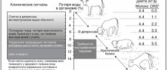
Professor I.P. Kondrakhin developed a test based on biochemical effects. He proposed precipitating coarse proteins using a solution of zinc sulfate. This test can determine the extent of the disease based on the amount of sediment. Indeed, as inflammation increases, the amount of proteins in the blood serum and, accordingly, sediment changes proportionally. Test readings are recognized as follows:
- in a healthy calf up to three months, the test shows 1.6–1.8 ml;
- with a mild or moderate course of the disease, the indicator is 1.5–1.3 ml;
- the maximum indicator of severe disease is 1.2 ml;
- if the test shows 0.9–0.8 ml, then the calf is on the verge of death.
Symptoms of bronchopneumonia
The development of catarrhal bronchopneumonia can occur in various forms of severity:
- In acute form.
- In subacute form.
- In chronic form.
Characteristics of the acute form of the disease
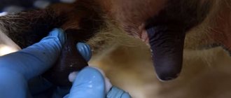
The acute form of the disease lasts from 5 to 10 days. During this period, the calf becomes lethargic, its appetite decreases, and slight malaise is noticeable. The animal breathes not through its nose, but through its open mouth. Hyperemia appears on the mucous membranes of the nose and eyes. The serous exudate that collects in the nose becomes purulent.
The animal’s health is quickly deteriorating, it is breathing harshly, moist wheezing is heard, the heart sounds are muffled, a blood test shows an increase in leukocytes. By the third day, the sick calf experiences a sharp increase in body temperature to 42 C. There is a deterioration in the general condition, the appearance of physical inactivity, rapid and difficult breathing is observed.
With bronchopneumonia, an increase in the concentration of leukocytes in the blood of the calf is observed
The acute course of bronchopneumonia changes the mucous membranes of most animals - they become noticeably pale. The upper respiratory tract swells, and the bronchi and bronchioles contain large amounts of exudate. Enlarged bronchial lymph nodes
Characteristics of the subacute form of the disease
The main symptoms of the subacute form of the disease are decreased appetite and decreased nutritional status within a month, and malnutrition develops. This phase is accompanied by an evening increase in body temperature by 1–2°C, the calf breathes with shortness of breath, accompanied by a wet cough. If the disease worsens, the condition may worsen: shortness of breath increases, hypoxia develops, digestion fails, and diarrhea appears. The animal becomes exhausted and a large amount of purulent mucus appears in the bronchi. The bronchi themselves are swollen, hyperemic, and hemorrhage is possible. The pleura contains a lot of fluid, the liver enlarges
Characteristics of the chronic form
A calf that has become ill with a chronic form of bronchopneumonia is significantly behind a healthy calf in growth and becomes hypotrophic. His cough does not stop, serous fluid flows from the nose, the mucous membranes become bluish, the body temperature rises slightly, and dry wheezing is heard in the lungs when listening. There is a constant change in appetite.
A healthy calf has pink mucous membranes and good weight
Course of the disease
The disease can occur in acute or chronic form. In particularly advanced cases, the course of the disease is accompanied by abundant formation of ulcers.
The acute form of pneumonia primarily involves a sharp increase in body temperature, coughing and discharge from the nose and eyes. The appetite and general condition of the animals are satisfactory, although apathy in the behavior of calves is sometimes observed. Sick animals do not move unless necessary and prefer to lie still. If the disease is started, its symptoms will begin to expand: the stool will change, the calves will begin to refuse food, etc.
The chronic course of pneumonia in cattle is characterized by a clear developmental delay:
- sick calves are smaller than their peers;
- there are bald spots and bald patches on the fur;
- dry skin.
Just as in the acute form, animals move little, cough and show constant shortness of breath. Occasionally, mucous nasal discharge can be observed in infected calves. The body temperature of sick calves is normal, which is one of the main differences between the chronic form of pneumonia and the acute form.
Important! The acute course of the disease most often occurs in calves 1-2 months old. The chronic form is more typical for older individuals - from 3 to 5 months.
Consequences
If treatment for pneumonia in cattle is started, it can become chronic or lead to serious disruptions in the functioning of the respiratory system. In addition, the cardiovascular and digestive systems, which are closely related to lung function, may be irreversibly damaged. Finally, if the animal is severely affected by the disease, it can subsequently cause a general weakening of the immune system in calves. They will generally be more susceptible to unfavorable environmental conditions and various infectious diseases.
Pneumonia is extremely rarely fatal.
Signs, symptoms and diagnosis
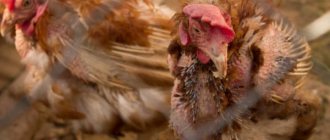
Bronchopneumonia in chickens can be recognized by the following symptoms:
- rapid breathing;
- wet rales;
- the activity of chickens sharply decreases, they sit all the time, cannot move independently, eat food, drink water;
- heavy breathing, the chicken breathes with its mouth open.
In the absence of appropriate therapy, young animals will begin to die on the 2nd day.
Attention! The disease can be detected in birds not only by signs, but also by living conditions.
To make a diagnosis, you do not need to resort to complex diagnostic methods. Bronchopneumonia can be diagnosed by external signs. Bioassays can confirm suspicions.
Stagnation of blood causes swelling
The pathogenesis of the disease catarrhal bronchopneumonia is quite complex. After all, almost all organs and vital systems of the calf are involved here. The disease strikes the first blow on the nervous system. Humoral and parallel nervous reactions are disrupted, which entails a decrease in the overall stability of the body.
In the blood of a calf, against the background of a sharp decrease in the percentage of histamine, the globulin protein fraction increases. This causes stagnation in the circulatory system and partial swelling of the mucous membrane throughout the entire area of the bronchi and bronchioles. Exudative processes and leukocyte reactions during the disease cause the accumulation of exudate simultaneously in both the bronchi and alveoli.
The lung tissue thickens and the calf first snorts and then coughs. Pathogenic and saprophytic microflora actively multiply, simultaneously releasing a lot of toxins into the body. Certain parts of the lung of a patient with bronchopneumonia cease to function normally, and this already leads to confused, rapid breathing and disruption of general gas exchange.
Treatment of pneumonia
Pneumonia in calves can only be treated by a veterinarian!
If such an attack occurs on your farm, it is better to immediately contact a veterinarian. Only a doctor can diagnose pneumonia and the exact cause of its occurrence (pathogen). Pneumonia can be successfully treated with antibiotics. However, the choice of one drug or another should be determined by the nature of the disease and the type of your calves (meat or milk production). Please note that when used on meat breeds, it is necessary to maintain a certain period before slaughter, since residual amounts of antibiotics can remain in the meat for a long time.
Intratracheal therapy
R. G. Mustakimov is considered the founder of intratracheal therapy. He recommends the use of intratracheal isoniazid at a dosage of 10 mg. In addition, the tetracycline group of antibiotics is used at a rate of 5000 units. per kg of calf weight. A 10 ml solution is made on the basis of novocaine (5%). You need to inject three times a day for six days.
To enhance the therapeutic effect and general strengthening of the animal during the disease with bronchopneumonia, trivitamin is injected intramuscularly into the calf, but only 2 mg should be injected once every three days. Also, to improve the overall picture, 80 ml of oxygen is administered twice per course to the abdominal cavity. The interval between administration is about four days.
It has been noticed that with oxygen, calves recover much faster. If this procedure is not available, then the entire course lasts nine days. Intratracheal treatment according to this scheme shows quite good results.