Liver diseases
There are a lot of liver diseases in cows. All these diseases can be divided into 3 main areas:
- diseases caused by viruses and other similar microorganisms;
- diseases caused by parasites (worms, worms, etc.);
- poisoning by toxins.
Since we are talking about non-contagious diseases of cattle, we are also interested in toxic poisoning. Not everyone knows, but hepatitis can be caused by long-term poisoning by toxins. It is not contagious, but in advanced acute form it can cause cirrhosis of the liver.
When this organ is damaged, the cow loses interest in everything, her appetite decreases, and a yellowish coating appears on the mucous membranes. In a relatively short time, the cattle loses a lot of weight, milk yield drops sharply, and milk may disappear completely.
Among non-contagious liver diseases of cows, hepatosis is common. This series of pathologies is caused by metabolic disorders in the body. Even newborn calves can suffer from hepatosis. Clinical manifestations are similar to those of hepatitis.
Conjunctivitis
Conjunctivitis is an inflammation of the eyes caused by viruses, bacteria or parasites. Sometimes it occurs as a result of injury and foreign particles entering the eyes. Chemical burns also cause conjunctivitis.
If the nature of the disease is viral, it is extremely contagious. In the absence of timely treatment, it leads to loss of vision in livestock.
Types of conjunctivitis:
- catarrhal - the cow's eye is half-closed or completely closed, swollen, profuse lacrimation, acute reaction to light.
- purulent - inflammation of the eyes with purulent discharge. The eyes become swollen and pus is released. Subsequently, ulcerations and abscesses form.
- phlegmonous - characterized by severe swelling and protrusion of the mucous membrane.
- follicular - determined by the proliferation of follicles in the third eyelid.
Treatment should begin at the first manifestations of the disease. It includes antibacterial therapy (intramuscular administration of the drug and local treatment of the mucous membrane).
Hyperacute form of PCH
Acute and subacute forms of the disease are identical in course. The only difference is that in the subacute form, symptoms appear a little later and death is delayed for a short time.
If measures are not taken to provide veterinary care, the infected animal dies within half a month.
When the hyperacute stage of malignant catarrhal fever occurs, the cattle die already on the 4th day. A number of other signs are added to the damage to the central nervous system:
- chills and long-term fever;
- cessation of lactation;
- complete refusal to eat;
- cardiopalmus;
- loose stool streaked with blood;
- strong thirst;
- difficulty breathing, sometimes attacks of suffocation;
- difficulty swallowing if the pharynx is affected;
- hot, dry nose.
There is a flow of mucus mixed with pus and blood from the animal's nostrils. Dead tissue also comes out. The discharge has a pungent smell of rot.
Such discharge is observed from the mouth.
Ulcerative formations can also appear on the cow’s genitals. The labia swell, the mucous membrane becomes inflamed and covered with ulcers. Sometimes a disorder of the genitourinary system appears. The pregnancy of pregnant cows ends in abortion.
It is important to monitor the body temperature of sick individuals. On the first day it rises sharply up to 42 degrees
On the second or third day it drops to 39-40 degrees. The temperature should remain at this level for some time. With a sharp decrease in body temperature, we can talk about the deterioration of the animal’s condition and the approach of death.
Diagnostic tests
If the symptoms of malignant catarrhal fever are similar to those of other diseases, a necessary condition for making a correct diagnosis are:
- laboratory research;
- analysis of general signs of infection;
- analysis of changes that occurred in the body of deceased individuals.
These studies must be carried out comprehensively, otherwise it will not be possible to obtain an accurate picture.
Using the above tests, the virus is detected. Often, tissue histology and examination of sections of altered cells are added to the mandatory research analyses.
After the death of the animal, pathological work is required to confirm or refute the diagnosis of malignant catarrhal fever. In order to officially approve this diagnosis, the following must be detected in the body of a deceased cow:
- protein particles on all mucous membranes;
- unhealthy lymph nodes;
- inflammation of the meninges;
- unnaturally thickened blood;
- enlarged spleen;
- many bruises in the ball of tissue;
- weak myocardium.
Diseases with similar symptoms include: rinderpest, foot and mouth disease, severe poisoning, rhinotracheitis, acute stool liquefaction, mycotoxin, listeriosis, rabies and others.
Leukemia
Leukemia is a chronic viral disease that affects the hematopoietic system. It affects weak cows with suppressed immunity. Individuals with good body resistance act as carriers.
Treatment does not bring results. If this disease is detected, blood tests of all animals are carried out, according to the results of which the cows are culled.
Symptoms appear at a late stage:
- Decreased appetite
- Anemia,
- Enlarged lymph nodes.
If these signs occur, the animal must be isolated until examined by a veterinarian.
Causes of hoof problems
In order to take adequate treatment measures, it is worthwhile to understand the causes of hoof disease in a cow as quickly as possible.
- Mechanical damage. It can be easily obtained when moving on hard, uneven ground.
- Quality of bedding in the stall. When putting weight on the leg, the emphasis is on the outer part of the hoof. On good, soft bedding, the weight of the cow is distributed evenly over the entire area of the legs; otherwise, the load falls only on the outer part. This causes hoof disease.
- Poor floor in the stall. Uneven floor coverings and protruding nails lead to injury.
- Neglect of care and maintenance conditions. These include high air humidity, infrequent manure removal, and dampness in the stall. Under the wrong conditions, existing microdamages can become infected and eventually develop a disease.
- Rare walks. In this case, deformation of the hooves occurs, since with a lack of movement, natural grinding does not occur. The animal begins to limp.
- Weak immune system. If the body is not able to protect itself from inflammation naturally, then the disease will progress. Typically, immunity weakens after illness or due to improper feeding of the cow.
Parasitic diseases
This type of disease is provoked by the appearance of parasites: lice, ticks, fleas. A cow can become infected with parasites from other animals, from humans, and when walking.
This group includes diseases such as: lice, trichomoniasis, thelaziosis, piroplasmosis. The main thing in treating these diseases is getting rid of parasites.
Functions of a book for cattle
The book is a section of the gastrointestinal tract, which is located between the mesh and the abomasum, on the right side. The volume of this compartment is about 15 liters. It grinds food and absorbs liquid from feed. They become dense and dry out.
The crushing of food masses occurs due to the fact that the mucous membrane of the book consists of folds that resemble leaves in a book. That's why this section was called a book.
There are a lot of leaves, and the contractions are quite weak, so the passage of food is slower. This is a normal process. Since food enters the book quickly and comes out slowly, food residues accumulate accordingly. This is all due to the fact that juices and liquids are thoroughly sucked out, leaving only dry scales and flakes. Gradually, dry food accumulates between the leaves, squeezes them, and food begins to spoil there and cause discomfort. Blood circulation is disrupted, and inflammatory processes begin in the body. This is how blockage occurs.
In calves, this department begins to work only when solid feed begins to be introduced into complementary feeding. When feeding with milk, blockages cannot happen, because liquid milk passes through the book unhindered.
Uterine diseases
The most dangerous of the non-contagious uterine diseases in cows is endometritis, in other words, inflammation of the uterine mucosa. The reasons may be different, but most often it is a difficult birth. The cause of endometritis can be abortion and even rough, unprofessional treatment of an animal during childbirth.
The disease can be acute, chronic and latent. If measures are not taken in time, endometritis will develop into either peritonitis or infertility. In the acute form, the cow's temperature rises, and there is constant discharge from the vagina.
Only a veterinarian can determine the latent and chronic forms. In this case, the animal can feel normal until the disease progresses to peritonitis. Therefore, in order to exclude a latent form of endometritis on days 8–10, tests must be taken in all cows after birth or abortion.
There are many more non-contagious cow diseases. We went through only the most frequently encountered ones. Share this information with your friends on social networks and perhaps your like will help identify a dangerous disease at an early stage.
Write a comment at the end of the article.
Cystitis
By definition, cystitis is inflammation of the bladder. It can occur in different forms.
The main symptoms include painful urination, especially at the last stage of the process. It happens that the disease is accompanied by false urges, the animal arches its back and tries to urinate, but as a result nothing happens or a few drops come out.
Laboratory tests reveal a high content of red blood cells in the urine. As a result, the urine becomes cloudy and pus may be released. But cystitis can only be a consequence of a more dangerous process.
Sometimes a cow becomes infected with some kind of sexually transmitted infection and, as it develops, symptoms similar to cystitis appear. Again, some forms of inflammation in the kidneys can be mistaken for cystitis.
The pathology is treated with antibiotics in combination with sulfonamides. Sometimes veterinarians prescribe washing the bladder with a solution of furatsilin in a ratio of 1:5000 or something similar. But all this is done after laboratory tests. Before taking the tests, you can only syringe the cow with furatsilin.
Smallpox in cattle
Cowpox is a viral disease that causes skin rashes, ulcers and pustules. Rashes or bumps can appear anywhere on the udder, stomach, leg, or torso. Smallpox is accompanied by a short-term and slight increase in the cow's body temperature. Lymph nodes may become inflamed if the general condition is unsatisfactory. Skin rashes may go away on their own after some time. The disease cannot be left untreated, since milk yield is significantly reduced due to the general condition of the cow.
If the cow's immune system is suppressed by medications or other diseases, the infection may progress to the next phase. Only a veterinarian can determine the presence of smallpox. Carriers of the disease in rural farms can be mice or even pets, so when working with an animal that has been diagnosed with smallpox, it is better to keep it in a separate pen for the duration of the disease. A special vaccine can be used to prevent smallpox. Before choosing a remedy, you should invite a veterinarian for a more detailed consultation; he will give an idea of the relevance of such an event.
In the room where cattle are kept and raised, there should be no dirt or bags of food for mice to flock to in herds. All holes in the barn must be sealed, and all pens and work equipment must be thoroughly cleaned every week using disinfectants. If the farm has special equipment for milking cows, they need to be washed and treated with antiviral solutions. Gloves should be worn when working with animals.
Reviews and
Mastitis
Mastitis in cows is an inflammation of the mammary glands, rendering milk unfit for consumption. Mastitis occurs as a result of stagnation of milk (with irregular milking of cows) and the presence of gynecological diseases leading to hormonal imbalance.
Symptoms:
- The udder is swollen.
- Pus comes out of the nipples.
- The milk is yellow.
- There are pronounced seals on the udder. The form is lost.
Treatment of inflammation occurs through intramuscular administration of antibacterial drugs.
Diseases of the reproductive organs
This group of diseases causes a decrease in the quality of milk
Therefore, it is important to promptly identify the disease and begin its treatment.
Non-infectious mastitis
The disorder is caused by poor living conditions for the animals. Mastitis is provoked by drafts, untimely change of bedding or its absence, lack of heating in the room, violations of the feeding regime, etc. The pathology is manifested by hardening or swelling of individual areas of the udder. If you milk an animal, curd-like clots are released into the milk, the smell of which is clearly unpleasant. Blood elements may appear.
During the treatment process, the cow must be milked frequently and carefully, especially the diseased part of the udder. The frequency of milking is 2-3 hours. This will require light massage procedures with rubbing of ointments prescribed by the doctor. To prevent the occurrence of mastitis, it is necessary to comply with the requirements for keeping and feeding animals.
Endometritis
Along with mastitis, this disease of the reproductive organs is a fairly common problem in cattle. Endometritis is associated with inflammatory processes in the area of the uterine mucosa. The disease is caused by complicated childbirth, which provokes injury to the mucous layer. The cause may be insufficiently careful obstetric care or a condition with retained placenta. Often, endometritis is a consequence of the animal’s low activity due to lack of walking, as well as insufficiently balanced feeding during pregnancy.
Symptoms are manifested by the appearance of cloudy exudate that has a gray or red tint. This happens abundantly 2-5 days after calving. If the cow is lying down, then the intensity and abundance of discharge increases. The animal is forced to arch its back or take a position as if urinating.
Udder swelling
The disease is manifested by thickening of the skin in the udder area, the size of which increases. The swelling gradually spreads to the abdominal cavity located near the udder. The cause of the pathology is a violation of the diet and regimen of a pregnant cow or heifer.
During the treatment process, it is necessary to regularly milk the cow at least 6 times daily. Procedures with udder massage will also be required, after which it will be necessary to treat it with camphor oil. During this period, the cow should not be fed concentrated feed; she can return to it only after recovery. To avoid pathology, you must:
- adhere to a balanced diet and proper feeding regimen;
- exclude feeding with succulent food 7-14 days before calving.
Pathologies of the digestive system and oral cavity
Ailments in the gastrointestinal tract are varied. Their risks increase when using low-quality feed or due to non-compliance with the diet.
Blockage in the esophagus
When a problem occurs, the animal becomes restless due to swallowing large food items. The cow begins to stretch her neck and lower her head
Noteworthy is the frightened appearance and active secretion of saliva when the scar is swollen. After some time, the heartbeat and breathing become more frequent, symptoms of suffocation occur, the mucous membranes of the eyes and mouth become cyanotic
These signs appear within a few hours. Without assistance, death is possible.
The pathology is caused by haste in the process of eating, as a result of which the food clogs the esophagus. The same result can be provoked by fear or stress. It is necessary to quickly remove the blockage, freeing the esophagus from food. If the upper segments of the esophagus are blocked, it is possible to push the product through with your hand. But in case of deep blockage, you will need to use a hose with a diameter of 30-40 mm. Before manipulation, it is necessary to pour up to 1 cup of vegetable oil into the esophagus area, which will ensure the desired degree of gliding. After eliminating the problem, feeding is excluded for 11-12 hours. As a preventive measure, it is advisable to grind the food.
Tympany
The pathology of scar swelling is accompanied by an increase in the volume of the left sector of the abdomen. The animal stops eating, its chewing gum disappears, and anxiety arises. At the next stage, the respiratory function becomes more frequent, the cow actively sweats, and becomes lethargic. Blueness appears on the mucous membranes of the oral cavity and organs of vision, and at the same time the ears and limbs become cold.
The development of pathology is provoked by failures or cessation of gas belching in case of overeating of feed with an easily fermentable effect. This applies to clover, alfalfa, and sainfoin. The problem also occurs after eating raw grass or food that has turned sour or fermented.
To eliminate the violation, it is necessary to remove gases from the rumen using a rubber hose with a diameter of 30-40 mm. Then a 4% solution of formalin in a volume of 1 liter or 2% ichthyol in a solution in a volume of 1/2 liter is injected into the scar. As a drink, you should use a solution of magnesia in the proportion of 35 g: 1 l. If the effect is not achieved, the veterinarian will have to puncture the scar
It is important to remember that it is easier to prevent a problem from occurring. Therefore, it is necessary to limit the animal’s consumption of easily fermented food and completely eliminate sour foods from the diet.
Stomatitis
The disease is accompanied by swelling of the oral mucosa, which becomes red and overly sensitive. The animal actively produces saliva. It cannot eat roughage, preferring to drink cool water, which relieves pain.
The reason is the excessively frequent consumption of dry type or solid food. Stomatitis is also caused by the consumption of herbs that irritate the mucous membranes or contain toxic components. In some cases, it becomes the first sign of the development of foot and mouth disease and some other ailments of an infectious nature.
During the treatment, the mouth is rinsed:
- 1% boric acid solution;
- 0.1% solution of potassium permanganate;
- 2% baking soda solution.
When ulcers appear on the mucous membranes of the mouth, they are treated with a 1% solution of copper sulfate or a 2% solution with silver nitrate. To eliminate the risk of developing stomatitis, limit feeding using roughage, and carefully select pastures.
Process of obtaining milk
Milk formation is affected by the quantity and quality of food received. It is clear that nutrients come from the blood, but they enter it through the digestive system.
Important
Nerve plexuses also play an important role in milk formation. Due to poor care and stress, the animal will reduce its milk yield. If a cow has illnesses, then after calving a hard udder is observed.
The rate of fluid filling is related to the hormone oxytocin. Under its influence, the cells of the alveoli contract, the fluid from them enters the nipples. The hormone appears in the blood due to massage and washing. The optimal duration of exposure is 4-5 minutes, after which the movement of milk stops. If milking is not quick, they cannot be expressed completely.
During the process, milkmaid operators are required to observe hygiene rules.
- Separate dishes are recommended for the first servings.
- The liquid may contain microorganisms that are found on the surface. To eliminate them, you need to pass liquid through the fabric for a bactericidal effect.
- Workers are required to wear clean bandages, scarves, and gloves.
- Persons working with milk undergo mandatory medical examinations.
- To prevent bacterial contamination of the drink, farms periodically fight flies and rodents. For these purposes, chemical and biological agents are used.
Tympany - swelling of the mesh and scar
Often this is a summer disease, the causes may be:
- The cow eats a lot of young and juicy grass, especially a lot of clover and other vegetation of the legume family;
- Eating large amounts of damp grass with dew, or rotten beet tops;
- It often happens in calves when the calf greedily drinks its mother's milk.
Symptoms of tympania: severe bloating, which is immediately visible; The animal also exhibits hunger pitting, restlessness, and lack of chewing.
Treatment of tympany in cows:
The left and right halves of the abdomen are alternately rubbed with a self-made tourniquet of straw, or, if available, with a brush; Rubbing the abdomen with turpentine will help, while kneading in the area of the hungry pit with a fist; The cow is bridled, and the rope is pre-wetted in kerosene or salt water (a tablespoon of rock salt per glass of water), these procedures are carried out in order to cause the animal to burp. It is important to try to get the animal to constantly move; movement will help cause belching.
When carrying out these measures, observe the behavior of the animal; if they do not help, then immediately contact a veterinarian, otherwise the animal may die.
Hernia
It may occur due to mechanical trauma due to negligence or due to congenital pathology.
Symptoms: a protrusion forms in the navel area, the calf refuses to eat, becomes nervous and excited, diarrhea occurs, and pain in the area of the tumor occurs.
Treatment: a specialist repairs the hernia with his hands at home. If this does not help, the animal is sent for surgical procedures.
Prevention: constant monitoring of young calves will help identify a hernia in the early stages of the development of pathology and relieve subsequent serious complications.
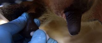
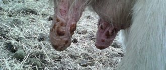
foot and mouth disease
Foot and mouth disease is an acute infectious disease that mainly affects ruminants, but can also spread to humans. The first two weeks the disease occurs latently, after which its main symptoms begin to appear (Figure 10):
- Bubbles with liquid inside form on the udder, mucous membranes and between the hooves;
- After a few days, the virus spreads through the blood and the animal begins to develop a fever (the temperature rises to 42 degrees);
- Secondary blisters appear, which are accompanied by normalization of temperature and profuse salivation;
- Bubbles between the hooves make it difficult for the animal to walk, the cow begins to limp or refuses to get up;
- When the blisters burst, ulcers form in their place, which burst within a few days.
Figure 10. Symptoms of foot and mouth disease in cows
Treatment of foot and mouth disease in cows should only be carried out by a veterinarian. Therefore, after discovering bubbles, you should immediately contact a specialist, isolate the sick animal and not let it out to pasture. Milk from a cow with foot-and-mouth disease can be eaten only after preliminary boiling or pasteurization. Meat can be used in food only after thorough heat treatment, but it is not allowed for sale.
Hematuria
Chronic hematuria in cattle is perhaps the most mysterious disease of our time. The etiology of this scourge is still not fully understood. For a long time it was believed that cows tend to become infected with hematuria on pastures when the animals are overcrowded. Accordingly, they looked for infection.
Then they looked for a certain genetic predisposition of a certain breed of cows to this disease. Now we have settled on the fact that the disease manifests itself geographically, that is, only in a certain area. Local animals become ill at the age of 2–3 years. Imported cows have approximately the same period of 2–3 years from the moment of transportation.
The visible symptoms are in many ways similar to genitourinary diseases and infections, for example, cystitis. The walls of the bladder and the cortex of the kidneys are affected; anemia can be diagnosed in other organs.
Modern veterinary medicine does not know why such diseases occur in cows. There is currently no clear course of treatment for hematuria. The disease can last from 2 months to several years, but invariably ends in the death of the cow.
foot and mouth disease
Symptoms
This disease of bulls and cows is dangerous to humans. Accompanied by an increase in body temperature and refusal to eat. In heifers, milk yield decreases, redness appears in the area of the mucous membranes, which indicates the beginning of an inflammatory process in the body. After some time, formations in the form of ulcers appear in the mouth.
The cows also experience an increase in salivation, and they begin to grind their teeth. Swelling often appears in the hoof area. If foot and mouth disease is not treated in a timely manner, the animal’s breathing becomes difficult, the nasopharynx is damaged and the nipples are modified.
Treatment
To treat foot and mouth disease, antibiotics are used, which must be prescribed by a doctor. In parallel with antibiotics, antiseptic drugs are used - they help heal wounds on the body that appear during foot and mouth disease. If you treat foot and mouth disease in a timely manner, you will be able to stop the disease and save the lives of the entire livestock.
Tuberculosis
Unfortunately, this disease is highly contagious for both animals and humans. A person can become infected with it from an animal either through the air or by consuming milk and meat from sick animals. Tuberculosis in cows is incurable, so sick animals are sent to slaughter. Characteristic signs in sick animals rarely appear.
Basically, the disease is asymptomatic and chronic. Sick animals are practically no different from healthy ones. Only sometimes, in very severe cases, the lungs of animals are affected and a rare but hysterical cough appears. Occasionally the temperature rises. Gradually the cough becomes weaker, but more painful.
The cow begins to breathe frequently, the mucous membranes of the mouth become bluish. Lymph nodes enlarge at the back above the udder, i.e. bumps appear that are hard and hot to the touch. To protect yourself and your loved ones from this terrible disease, you just need to examine your cows twice a year during scheduled treatments.
This disease is classified as infectious.
The causative agent of tuberculosis is mycobacterium, of various types (human, bovine and avian).
This disease is transmitted through already infected animals by airborne droplets, as well as through contact with affected areas of the mucous membrane. Transmission can occur through shared feed, manure, water, bedding, and shared care items.
Tuberculosis is not noticeable externally, since it does not produce symptoms. This disease mainly affects the lungs and intestines.
We suggest you familiarize yourself with: Wirehaired Fox Terrier, character, diseases, care, photos
If tuberculosis has affected the lungs, the animal begins to cough violently, which indicates damage to the pleura and the lungs themselves. If the intestines are affected, the cow will experience bloody diarrhea.
With widespread damage, animals become very thin and weakened, their lymph nodes become enlarged, and they eat little.
To detect the presence of Koch's bacillus in an animal, you need to submit bronchial mucus, milk, urine or feces for analysis. If an animal is infected, it must be destroyed in order to prevent infection of healthy cows. Regular intradermal tuberculinization of all livestock is used as preventive measures.
Preventive actions
Any disease is much easier to prevent than to treat.
Here is a set of preventive measures to protect livestock from bursitis of any kind:
Grazing areas. If you have a choice, you should not allow cows to graze on rocky terrain.
It is worth paying attention to the plants - they should not be prickly. Warm. The barn should be warm, but don’t forget about regular ventilation
If the floor is cold, comfortable bedding is a must. Hygiene. Regular cleaning is key to the health and safety of livestock. Feeders. When designing a room for keeping cows, take into account their dimensions so that they do not injure each other while eating. Transportation. If you need to transport your herd, do so strictly following transportation rules to ensure the safety of the animals.
In addition, regular examinations by a veterinarian will allow timely detection of the development of any diseases. By following these rules, you can protect not only cows, but also any type of cattle from bursitis. And if you have already encountered the disease, you will be fully equipped to combat it.